


From the article you will learn the characteristics of small pelvic varicose veins in women - this is a deformed form of the veins of the pelvis with impaired blood flow in the internal genital organs andoutside.
general information
In the medical literature, small pelvic varices are also known as "pelvic congestion syndrome", "varicocele in women", "chronic pelvic pain syndrome". The prevalence of small pelvic varices increases with age: from 19. 4% in girls younger than 17 years to 80% in premenopausal women. Most often, pathology of the iliac veins is diagnosed during the reproductive period in patients aged 25-45 years.
In the majority of cases (80%), varicose veins affect the ovarian veins and extremely rarely (1%) are observed in the veins of the broad ligament of the uterus. According to the approach of modern medicine, the treatment of VTE should not be done so much from the point of view of obstetrics and gynecology, but first of all, from the point of view of veins.
Pathological Triggers
According to varicose veins of the pelvic organs in women, doctors understand the structural changes of the vessel walls that are characteristic of other diseases - weakening, followed by dilation and the formation of "vesicles". "The inside causes the blood to stagnate. Cases where only the vessels of the pelvic organs are affected are extremely rare. In about 80% of patients, along with this form there are signs of varicose veins in the groin, veins of the lower extremities.
The prevalence of varicose veins of the small pelvis is most pronounced in women. This is due to anatomical and physiological features, which show a tendency to weaken the venous walls:
- hormonal fluctuations, including those related to the menstrual cycle and pregnancy;
- increased pressure in the small pelvis, which is typical for pregnancy;
- more active periods fill the veins with blood, including the menstrual cycle, during pregnancy, as well as during sex.
All of these phenomena belong to the category of factors that provoke varicose veins. And they are found only in women. The greatest number of patients face varicose veins of the small pelvis during pregnancy, since there is a simultaneous distribution of provoking factors. According to statistics, in men, varicose veins of the small pelvis are 7 times less common than in men of the normal sex. They have a more diverse range of triggers:
- hypodynamia - long-term maintenance of low physical activity;
- increase physical activity, especially weightlifting;
- fat;
- lack of enough fiber in the diet;
- inflammatory processes in the organs of the genitourinary system;
- sexual dysfunction or refusal to have sexual intercourse.
A genetic predisposition can also lead to pathology of the plexus located inside the small pelvis. Statistically, varicose veins of the perineum and pelvic organs are most commonly diagnosed in women who have a close relative with the condition. The first changes in surname can be observed in adolescence during puberty.
The greatest risk of developing inguinal varicose veins in women is related to the pelvic blood vessels observed in patients with venous disease in other parts of the body. In this case, we are talking about congenital weakening of the veins.
Pathogenesis
Obstetricians and gynecologists believe that the following main reasons always contribute to the occurrence of VVP: valve failure, venous obstruction, and hormonal changes.
Pelvic venous obstruction syndrome can develop from congenital absence or absence of venous valves, this has been revealed by anatomical studies in the last century, and modern data confirm this. .
It has also been found that in 50% of patients, varicose veins are hereditary. FOXC2 was one of the first genes identified to play an important role in the development of VVP. At present, the relationship between disease development and mutations in genes (TIE2, NOTCH3), thrombomodulin levels and transforming growth factor type 2 β has been determined. These factors contribute to changes in the structure of the valve itself or the wall of the vein - all this leads to the failure of the valve structure; enlarged veins, causing a change in valve function; to progressive reflux and eventually varicose veins.
An important role in the development of the disease can be played by connective tissue dysplasia, the morphological basis of which is a decrease in the content of different types of collagen or a violation of the ratio between them, leading todecreased venous strength. .
The incidence of VVP is proportional to the amount of hormonal changes, which are especially pronounced during pregnancy. In pregnant women, the capacity of the pelvic veins is increased by 60% due to the mechanical stress of the pelvic vessels and the vasodilating effect of progesterone. This varicose vein condition persists for a month after birth and can cause venous valve failure. In addition, during pregnancy, the volume of the uterus increases, a change in its position occurs, dilation of the ovarian veins, followed by venous obstruction.
Risk factors also include endometriosis and other inflammatory diseases of the female reproductive system, estrogen therapy, adverse working conditions for pregnant women, including heavy physical labour. prolonged labor and forced posture (sitting or standing) during the working day.
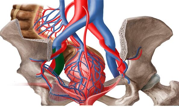
The formation of veins in the small pelvis is also facilitated by the anatomical features of outflow from the veins of the small pelvis. The diameter of the ovarian veins is usually 3 to 4 mm. The long, thin ovarian vein on the left empties into the left renal vein, and the right empties into the inferior vena cava. Normally, the left renal vein lies anterior to the aorta and posterior to the superior mesenteric artery. The physiological angle between the aorta and the superior mesenteric artery is approximately 90°.
This normal anatomical position prevents compression of the left renal vein. On average, the angle between the aorta and the superior mesentery in adults was 51 ± 25°, in children - 45. 8 ± 18. 2° in boys and 45. 3 ± 21. 6° in girls. In the case of an angle reduction from 39. 3 ± 4. 3° to 14. 5°, aortic mesenteric compression, or ganglion fissure syndrome, occurs. This is the so-called anterior chestnut syndrome, or rightly, of the greatest clinical significance. Posterior ganglion fissure syndrome has occurred in rare cases in patients with posterior aortic or annular arrangements of the distal left renal vein. Obstruction of the proximal venous bed causes an increase in pressure in the renal vein, which leads to the formation of retrograde reflux in the ovarian vein contrary to the development of chronic iliac venous insufficiency.
May-Turner syndrome - compression of the left iliac vein by the right iliac artery - is also one of the etiological factors of iliac veins. It occurs in no more than 3% of cases, it is found more often in women. Currently, due to the advent of radiotherapy and endovascular methods, this disease is more and more detected.
Classify
Varicose veins are divided into the following types:
- Primary type of varicose veins: an increase in the blood vessels of the pelvis. There are two types of valvular heart failure: acquired or congenital.
- Secondary thickening of the iliac veins is diagnosed exclusively in the presence of gynecological pathology (endometriosis, cancer, polycystic).
Varicose veins of the pelvis develop gradually. In medical practice, there are several main stages in the development of the disease. They will be different depending on the presence of complications and the spread of the disease:
- First level. Changes in the structure of the ovarian venous valve can occur for genetic or acquired reasons. The disease is characterized by an increase in the diameter of the veins up to 5 mm. The left ovary has marked enlargement in the external parts.
- Second level. This degree is characterized by the spread of pathology and damage to the left ovary. The veins in the uterus and right ovary may also become dilated. Expansion diameter reaches 10 mm.
- Third level. The diameter of the veins increases to 1 cm, the dilation of the veins is observed in the right and left ovaries equally. This stage is due to pathological phenomena of a gynecological nature.
It is also possible to classify the disease depending on the main cause of its development. There is a primary degree, where dilation is due to defective venous valve function, and a secondary degree, a consequence of female chronic diseases, inflammatory processes or complications of a natural nature. cancer. The extent of the disease may vary according to the anatomical features, indicating the location of the vascular disorder:
- Many classes.
- Vulva and perineum.
- Combined forms.
Clinical Manifestations and Symptoms
In women, pelvic varices are accompanied by severe, but nonspecific, symptoms. Usually, the manifestations of this disease are considered signs of gynecological disorders. The main clinical symptoms of groin varices in women that involve the pelvic blood vessels are:
- Abdominal pain without menstruation. Their intensity depends on the stage of venous damage and the extent of the process. For grade 1 of small pelvic varices, mild cyclic pain extending to the lower back is characteristic. In the later stages, pain in the abdomen, perineum and lower back, is persistent and intense.
- Mucus discharge. The so-called leucorrhoea has no unpleasant odor, does not change color, which means infected. The discharge volume increases during the second phase of the cycle.
- Increased symptoms of premenstrual syndrome and dysmenorrhea. Even before menstruation begins, the pain in women increases, to the point of difficulty walking. During menstruation, menstrual bleeding can become unbearable, spreading to the entire pelvic area, perineum, lower back, and even down to the thighs.
- Another characteristic sign of varicose veins in the groin in women is discomfort during sex. It is palpable in the vulva and vagina and is characterized by a dull ache. It can be observed at the end of intercourse. In addition, the disease is accompanied by increased anxiety, irritability, mood swings.
- Just like varicose veins of the small pelvis in men, in women of patients with such a diagnosis, interest in sex gradually disappears. The cause of the dysfunction is both constant discomfort and a decrease in the production of sex hormones. In some cases, infertility can occur.
Diagnostic tools
The diagnosis and treatment of varicose veins is carried out by a phlebologist, a vascular surgeon. Currently, the number of VVP detection cases is increasing due to new technology. Patients with CPP are examined in several stages.
- The first stage is a routine examination by a gynecologist: history examination, manual examination, ultrasound of the pelvic organs (to rule out other pathologies). Based on the results, an additional examination is indicated by the proctologist, urologist, neurologist and other relevant specialists.
- If the diagnosis is unclear, but VVPT is suspected, in stage two, angioscanning ultrasound (USAS) of the iliac veins is performed. This is a non-invasive, highly informative diagnostic and screening method that is applicable to all women with suspected VVPT. If in the past it was believed that just examining the pelvic organs was enough (venous examination was considered inaccessible and optional), in the present stage, pelvic venous ultrasound is the procedure. compulsory examination. With the help of this method, it is possible to determine the presence of varicose veins of the small pelvis by measuring the diameter, velocity of the blood flow in the veins, and preliminary to find out the pathogenesis of the disease. early - failure of the ovarian veins or venous obstruction. In addition, this method is used for dynamic evaluation of conservative and surgical treatment of VVPT.
- The study was performed vaginally and trans-abdominal. The veins of the parameter, the inguinal plexus, and the uterine veins were visualized vaginally. According to different authors, the diameters of the vessels of the named regions ranged from 2. 0 to 5. 0 mm (mean was 3. 9 ± 0. 5 mm), i. e. no more than 5 mmand the mean diameter of the arc ridges was 1. 1 ± 0. 4 mm. Veins larger than 5 mm in diameter are considered dilated. The inferior vena cava, iliac vein, left renal vein, and ovarian vein were examined transabdominal to rule out thrombosis and extravascular compression. The length of the left renal vein is 6 to 10 mm and its average width is 4 to 5 mm. Normally, the left renal vein where it crosses the aorta is slightly flattened, but its 2–2. 5-fold decrease in transverse diameter occurs without a significant acceleration of blood flow, which ensuresnormal flow without increasing pressure. region. In the case of venous stenosis due to pathological compression, its diameter is significantly reduced - 3. 5–4 times, and blood flow acceleration - above 100 cm / s. The sensitivity and specificity of this method were 78 and 100%, respectively.
- Ovarian vein examination is part of the mandatory procedure for pelvic vein examination. They are located along the anterior abdominal wall, along the rectus abdominis muscle, slightly toward the ileal veins and arteries. A sign of ovarian venous insufficiency in USAS is considered to be more than 5 mm in diameter in the presence of retrograde blood flow. In order to fully examine, prevent recurrence, and treat with the right strategy, an ultrasound scan of the veins of the lower extremities, perineum, vulva, inner thighs, and buttocks must be performed.
- The development of medical technology has led to the use of new diagnostic methods. In the third stage, after confirming the diagnosis by ultrasound, radiation diagnostic methods are used to confirm it.
- Pelvic angiography with bilateral selective contrast ovary is one of the radiologically invasive diagnostic methods performed only in the hospital. This method has long been considered the diagnostic "gold standard" to assess dilatation and detect iliac valve insufficiency. The essence of the method is to deliver contrast under X-ray control through a catheter inserted into one of the major veins (carotid, iliac or femoral) to the iliac veins. , kidneys and ovaries. Thanks to this, it is possible to identify anatomical variations of the structure of the ovarian veins, determine the diameter of the genital and pelvic veins.
- The retrograde contrast of the gonadal veins at the height of the Valsalva maneuver serves as a pathological angiographic marker of their valvular insufficiency with marked dilatation and corresponding curvature. . It is the most accurate method for detecting May-Turner syndrome, post-thrombotic changes in the thorax and inferior vena cava.
- When the left renal vein is compressed, adrenocortical vasculature with retrograde blood flow into the renal vein, contrast media stasis in the renal vein is identified. Measurement of the pressure gradient between the left kidney and the inferior vena cava. Usually, it is 1 mm Hg. Art . ; gradient equal to 2 mm Hg. Art. , Can suggest light compression; with gradient>3 mm Hg. Art. can be diagnosed with aortic-mesenteric compression syndrome with hypertension in the left renal vein, and gradient>5 mm Hg. Art. considered a hemodynamically significant stenosis of the left renal vein. Determination of the pressure gradient is an important element of the diagnosis, since, depending on its values, essentially different surgical interventions on the veins of the small pelvis are planned, whichvery important in modern conditions. Currently, this study (with normal pressure gradient) can be used for a therapeutic purpose - for ovarian venous thrombosis.
- The radiation method followed by computed tomography of the iliac veins with labeled red blood cells in vitro. It is characterized by the deposition of labeled red blood cells in the veins of the pelvis and visualization of the jugular veins, allowing identification of the venous plexus of the small pelvis and dilated ovarian veins. In different locations, the degree of iliac vein obstruction, reflux of blood from the iliac veins into the hemispheric veins of the legs and perineum. Normally, the ovarian veins were unenhanced, and no radiopharmaceutical accumulation was observed in the venous plexus. To objectively assess the degree of small iliac vein obstruction, the iliac vein obstruction coefficient is calculated. But this method also has the disadvantages of being invasive, relatively low spatial resolution, cannot accurately determine the diameter of the vein, so it is not commonly used in clinics today.
- Video endoscopic examination is a valuable tool for evaluating undiagnosed cases. Combined with other methods, it can help determine the cause of pain and prescribe the correct treatment. With varicose veins of the small pelvis in the ovarian region, along the round and broad ligaments of the uterus, the veins can be visualized as cyanotic, dilated vessels with thin walls and stretch. The use of this method is significantly limited by the following factors: presence of retroperitoneal fatty tissue, ability to assess varicose veins in a limited area, and inability to identifyvenous reflux. The use of this method is now diagnostically justified in suspected cases of multifocal pain. Laparoscopy allows visualization of causes of CPP, for example, foci of endometriosis or adhesions, in 66% of cases.
Features of therapy
For the definitive treatment of varicose veins of the small pelvis, a woman must follow all the recommendations of the doctor, and also change her lifestyle. First of all, you need to pay attention to the load, if it is too high, it must be reduced, if the patient has an excessively sedentary lifestyle, it is necessary to play sports, walk more often, etc. v.
Patients with varicose veins are recommended to adjust their diet, consuming as little junk food as possible (fried, smoked, sweet in bulk, salty, etc. ), alcohol, caffeine. It is better to give preference to vegetables and fruits, dairy products, cereals.
In addition, for prevention of disease progression and for medicinal purposes, doctors prescribe patients with varicose veins to wear compression underwear.
Drugs
ERCT therapy implies several important points:
- get rid of the reverse flow of venous blood;
- reduce the symptoms of the disease;
- stabilizes vascular tone;
- Improves blood circulation in tissues.
Preparations for varicose veins should be taken in courses. The remaining drugs, acting as pain relievers, are only allowed to be taken when in pain. For effective treatment, doctors often prescribe the following drugs:
- phleboprotectors;
- enzyme preparations;
- drugs that reduce the inflammatory process with varicose veins;
- Medicines to improve blood circulation.
Surgical treatment
It is worth recognizing that conservative treatments give truly visible results mainly in the early stages of varicose veins. At the same time, the problem can be basically solved, and the disease can be completely eliminated only by surgical methods. In modern medicine, there are many surgical methods of treating varicose veins, let's consider the most popular and effective types of surgery:
- thrombosis of the veins in the ovary;
- therapeutic therapy;
- resin of the uterine ligaments;
- laparoscopic removal of hypertrophic veins;
- clamp the veins in the small pelvis with special medical forceps (cutting);
- transverse - venous ligation (indicated if, in addition to the pelvic organs, the vessels of the lower extremities are affected).
During pregnancy, only symptomatic treatment of small pelvic varices is possible. We recommend wearing tights, taking blood tonics as recommended by the vascular surgeon. In the II-III trimesters, perineal vein atherosclerosis can be performed. If, due to varicose veins, there is a high risk of bleeding during spontaneous delivery, the choice is made in favor of a cesarean section.
Physical therapy
The physical activity system for the treatment of varicose veins in women includes exercises:
- "Bike". We lie on our backs, with our hands behind our heads or along our bodies. Lifting our legs, we make circular movements with them, as if we were riding a bicycle.
- "Aries". We sit face down on any hard and comfortable surface. Lift your legs up and gently launch them behind your head. Using your hands to support your waist and elbows on the floor, slowly straighten your legs and lift your body up.
- "Scissors". The starting position is on the back. Raise your closed legs slightly above the floor. We spread the lower limbs to the sides, bring them back and repeat.
Possible complications
Why are small iliac veins dangerous? The following consequences of the disease are often noted:
- inflammation of the uterus, its appendages;
- uterine bleeding;
- abnormalities in the work of the bladder;
- the formation of venous thrombosis (a small percentage).
Preventive
In order for varicose veins in the small pelvis to disappear as soon as possible and in the future not to relapse the pathology of the pelvic organs, it is necessary to observe simple rules of prevention:
- perform daily physical exercises;
- prevent constipation;
- adhere to a diet in which there must be vegetable fiber;
- not stay in one position for a long time;
- perineal contrast bath;
- so that varicose veins do not appear, it is better to wear shoes and especially comfortable clothes.
Preventive measures aimed at reducing the risk of onset and progression of varicose veins in the small pelvis are primarily about reducing normalcy of lifestyle.